Liver cancer is the third leading cause of death worldwide, and hepatitis B virus (HBV) infection has been identified as a major risk factor for its development. The occurrence of HBV-associated hepatocellular carcinoma (HCC) is attributed to multiple mechanisms, such as chronic inflammation and hepatocyte regeneration caused by a virus-induced cytotoxic immune response, abnormal activation of proto-oncogenes due to HBV DNA insertion mutations, and epigenetic alterations mediated by viral oncoproteins. The HBV envelope protein, hepatitis B surface antigen (HBsAg), is a key indicator of the increased risk of developing HCC in HBsAg-positive individuals. HBsAg is essential throughout the entire tumor development process, from initiation to progression, and as an oncoprotein, it participates in accelerating tumor progression.
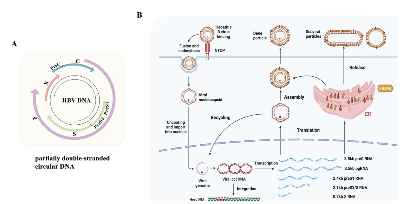
(Data source: Hao B, et al. Exp Hematol Oncol. 2025)
The structure and receptors of HBV and HBsAg
Hepatitis B virus (HBV) is a DNA virus belonging to the Hepatoviridae family. Under an electron microscope, a complete HBV particle exhibits a spherical morphology and consists of two distinct parts: an outer shell and a core. The outer shell is composed of hepatitis B surface antigen (HBsAg), glycoproteins, and lipids, while the core mainly contains the core protein (HBcAg) and the HBV genome.
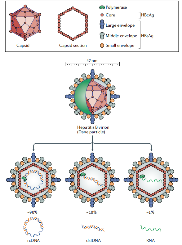
(Data source: Iannacone M, et al. Nat Rev Immunol. 2022)
The HBV genome consists of a partially double-stranded circular DNA molecule, ranging in length from 3182 to 3248 base pairs, depending on the genotype. This genome contains four functionally distinct open reading frames (ORFs) on the same strand: preC/C, X, P, and preS/S, which collectively encode seven polypeptides. The preC/C gene has two co-located start codons, encoding the viral pronuclear protein and core protein, respectively. The open reading frame of the pronuclear protein gene encodes a hydrophobic guide peptide that directs the pronuclear protein to the endoplasmic reticulum for cleavage, resulting in secreted HBeAg. The X gene encodes a 154-amino acid regulatory protein called HBx, which lacks DNA-binding activity and promotes oncogenesis through various mechanisms, including transcriptional activation. HBx is the most widely studied oncoprotein in HBV. The largest gene, the P region, completely overlaps with the envelope gene and partially overlaps with the preC/C and X genes. The protein encoded by the P region possesses key activities such as DNA polymerase, reverse transcriptase, and ribonuclease H, which are crucial for viral replication. The pre-S/S gene contains three in-frame start codons, resulting in the pre-S/S protein being divided into three distinct domains: Pre-S1, Pre-S2, and the S domain. These domains collectively encode three envelope proteins: the large envelope protein (LHB, 389-400 aa), the medium envelope protein (MHB, 281 aa), and the small envelope protein (SHB, 226 aa), collectively referred to as HBsAg. The Pre-S1, Pre-S2, and S domains together constitute LHB. The PreS1 domain interacts with the sodium taurine-cholic acid cotransport polypeptide (NTCP) receptor on the surface of hepatocytes, which is crucial for viral infection and entry into host cells.
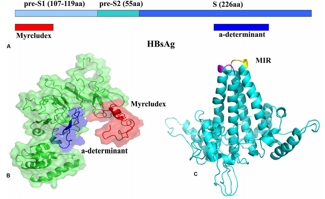
(Data source: Mobini S, et al. Front Immunol. 2020)
Carcinogenic effects of HBsAg
HBsAg induces HCC tumorigenesis by promoting proliferation. After HBV infects hepatocytes, the resulting envelope proteins induce more active cell proliferation through multiple intracellular signal transduction pathways, thereby promoting HCC tumorigenesis.
The Src/PI3K/Akt pathway is activated by proximal stimulation of the PKCα/Raf1 signaling pathway, inducing cell proliferation and tumor formation.
By increasing the expression of LEF-1 in the WNT signaling pathway, cyclin D1 and c-Myc are upregulated, which promotes more active cell proliferation and enhances the malignant transformation of hepatocytes.
by activating the NF-κB signaling pathway and upregulating long non-coding RNAs, which are known to be involved in inflammation-induced tumorigenesis.
It triggers PKC-dependent activation of the c-Raf-1/MEK/Erk 2 signaling pathway to regulate AP-1 and promote hepatocyte proliferation.
Inducing autophagy and NF-κB activation to promote cell proliferation, cell cycle transition from S phase to G2/M phase, and epithelial-mesenchymal transition to promote HCC progression.
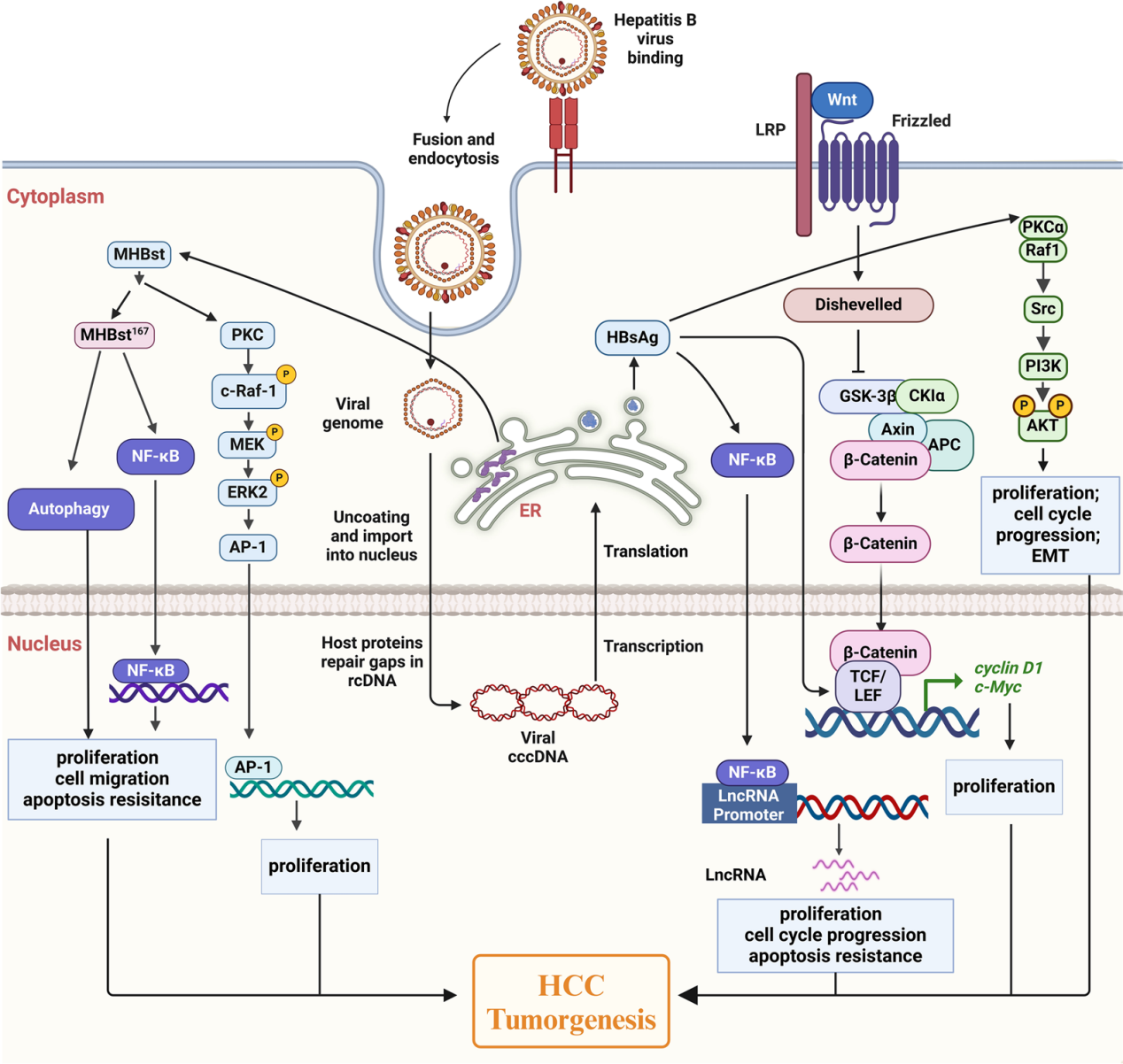
(Data source: Hao B, et al. Exp Hematol Oncol. 2025)
Immunosuppressive effect of HBsAg
HBsAg suppresses the immune response by impairing the phenotype and function of immune cells. Normal immune function is essential for killing tumor cells. HBsAg impairs the immune system by inhibiting the function of monocytes, dendritic cells, NK cells, and macrophages, creating a suppressive tumor immune microenvironment that favors the progression of HBV-related HCC.
HBsAg is associated with dysfunction of plasmacytoid dendritic cells. It can suppress immune system responses and promote a microenvironment favorable for cancer progression by downregulating Toll -like receptor (TLR) expression and inhibiting TLR9-mediated plasmacytoid dendritic cell maturation, as well as suppressing the secretion of cytokines, including IL-12, TNF-α, IFN-α, IFN-λ1, and IFN-λ2.
Natural killer (NK) cells, as effector cells of the innate immune system, play a crucial role in defending against hepatitis B virus (HBV) infection. HBsAg has the potential to influence the phenotype and function of NK cells . In vitro studies have shown that HBsAg can inhibit the activation of NK cells, secretion of cytokines, and release of cytotoxic substances through signal transduction and phosphorylation of STAT1 (STAT1), and a negative correlation was observed between NK cell activation and cytotoxicity and HBsAg levels. Furthermore, the presence of HBsAg in HBV-infected HCC cells stimulates the release of cytokines, including IL-10, TNF-α, and TGF-β, from suppressor monocytes via the MyD88/NF-κB signaling pathway. Simultaneously, it also stimulates the high expression of the inhibitory molecules programmed cell death ligand 1 (PD-L1) and major histocompatibility complex-E (HLA-E) in suppressor monocytes, triggering an immunosuppressive cascade through the interaction of PD-L1/PD-1 and HLA-E/CD94, leading to the production of regulatory NK cells that release IL-10. These cells can promote HBV immune escape by inhibiting the activation of autologous NK cells and CD4+ and CD8+ T cells through an IL-10-dependent pathway.
Intrahepatic monocyte-derived macrophages also regulate antitumor immune responses. HBsAg can inhibit TLR2-induced phosphorylation of p38 and JNK/MAPK, leading to a reduction in pro-inflammatory cytokines such as IL-6, TNF-α, and IL-12 produced by human monocytes. Furthermore, in HCC cell lines expressing HBV DNA, HBsAg promotes the deacetylation of the Notch 1 intracellular domain by upregulating sirtuin 1 expression, resulting in M2 macrophage polarization and the induction of immune tolerance in HBV-infected tumor cells.
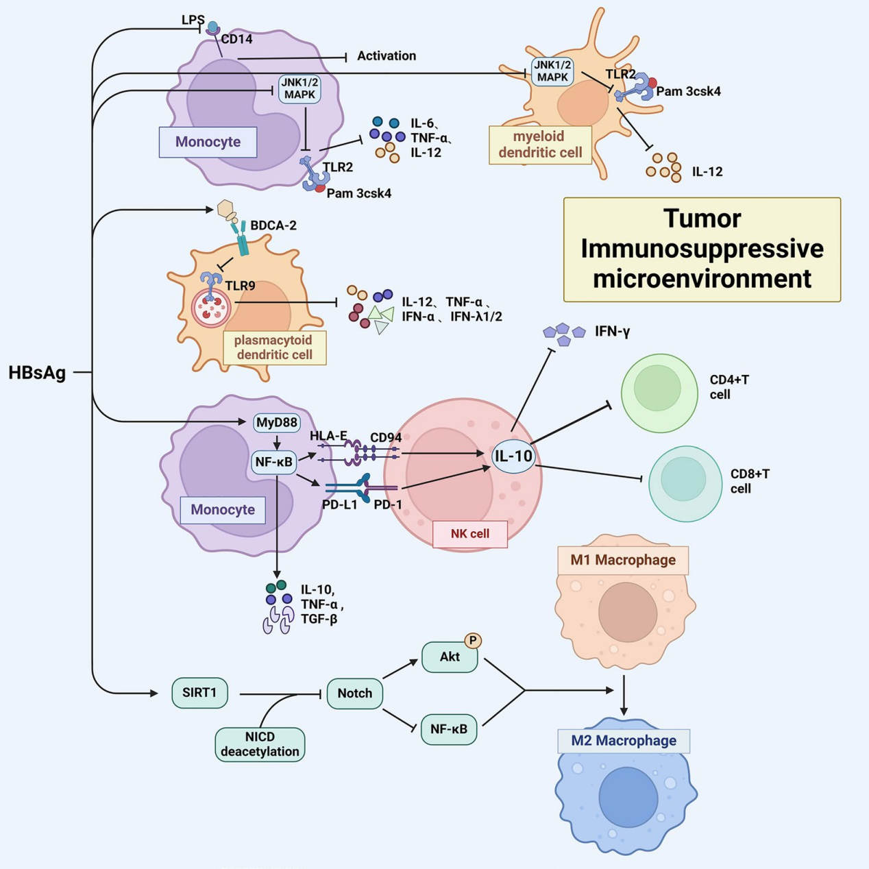
(Data source: Hao B, et al. Exp Hematol Oncol. 2025)
Targeted therapy for HBsAg
HBV infection has been identified as a major risk factor for over 80% of HCC cases. For HBV-related HCC patients, in addition to anti-tumor therapy, antiviral therapy is crucial for reducing HCC mortality and recurrence rates. Currently, clinically used antiviral drugs are mainly divided into two categories: nucleoside (acid) analogs and interferon drugs. Treatment methods currently in the experimental stage include entry inhibitors, RNA interference agents, capsid assembly modulators, and immunomodulatory methods. Entry inhibitors, as entry-blocking antiviral agents, competitively and irreversibly bind to the large envelope protein of HBsAg to NTCP, preventing HBV from entering host cells.
Libevitug (HH-003) is the first fully human monoclonal antibody targeting the PreS1 domain of HBV/HDV. Developed by Huahui Health based on fundamental discoveries about the NTCP receptor, Libevitug has received Breakthrough Therapy Designation (BTD) from both the NMPA and FDA. It was selected as “Best of EASL 2023–Viral Hepatitis”at the EASL Annual Meeting, and its pivotal registration clinical data (IIb) was presented at the AASLD 2025 Annual Meeting, gaining high recognition from the global academic community. Conditional approval for Libevitug is expected to be granted by the NMPA in January 2026, following the acceptance of its NDA in December 2024.
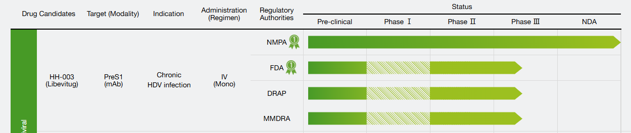
(Data source: Huahui Health official website)
Brelovitug (BJT-778) is an investigational, fully humanized monoclonal antibody for the treatment of chronic hepatitis B virus (HDV) infection, the most severe form of viral hepatitis. It is engineered to recognize and bind to a specific protein present on the surface of both hepatitis B and hepatitis C viruses. By binding to HBsAg, it clears HDV from the blood and prevents HDV from infecting new liver cells. By targeting this common protein, Brelovitug aims to help reduce the amount of virus in the body. Brelovitug has received Breakthrough Therapy Designation from the U.S. Food and Drug Administration (FDA) for the treatment of HDV and PRIME and Orphan Drug Designation from the European Medicines Agency (EMA).
In the phase 2b study of AZURE-1, the efficacy of Brelovitug in patients with HDV was evaluated. Virological response results at week 24 showed 100% response in the 300 mg weekly group and 75% in the 900 mg monthly group, while no cases were observed in the delayed treatment group (defined as a reduction of ≥2 log10 in HDV RNA from baseline or undetectable HDV RNA, i.e., <limit of detection, TND). The primary composite endpoints, virological response and normalization of alanine aminotransferase (ALT), were achieved in 45% and 35% of patients in the 300 mg and 900 mg groups, respectively, but not in the delayed treatment group. No treatment-related serious adverse events or treatment interruptions were observed in this study. The most common adverse event was injection site redness and swelling (14%–20%).
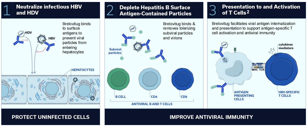
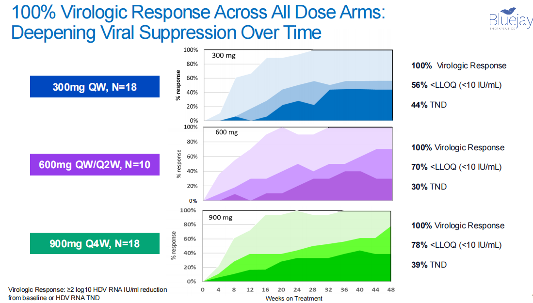
(Data source: AASLD 2025)
