Integrin α5β1 (ITGA5B1) is a key signaling protein between cells and the extracellular matrix, formed by the nonvalent binding of the α5 subunit (ITGA5, CD49e) and the β1 subunit (ITGB1, CD29). It plays a crucial role in biological processes such as cell adhesion, migration, differentiation, and angiogenesis. Integrin α5β1 has a significant role in bone formation and related diseases, and it is also an important mediator of tumor-stromal interactions.
Expression distribution of ITGA5B1
ITGA5 expression is cell-selective, primarily expressed in endothelial cells (vascular endothelial cells), fibroblasts, and a few epithelial cells. ITGB1 is widely expressed in the vast majority of cells, sharing a β subunit with multiple integrins, and is expressed in endothelial cells, mesenchymal cells, epithelial cells, and immune cells (T cells, B cells, monocytes/macrophages, platelets). Only when both are co-expressed does it constitute the functional integrin α5β1.
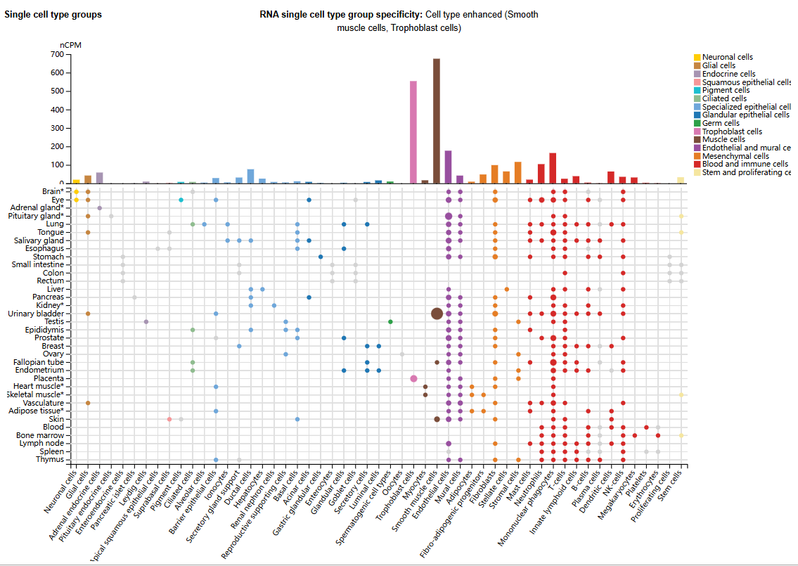
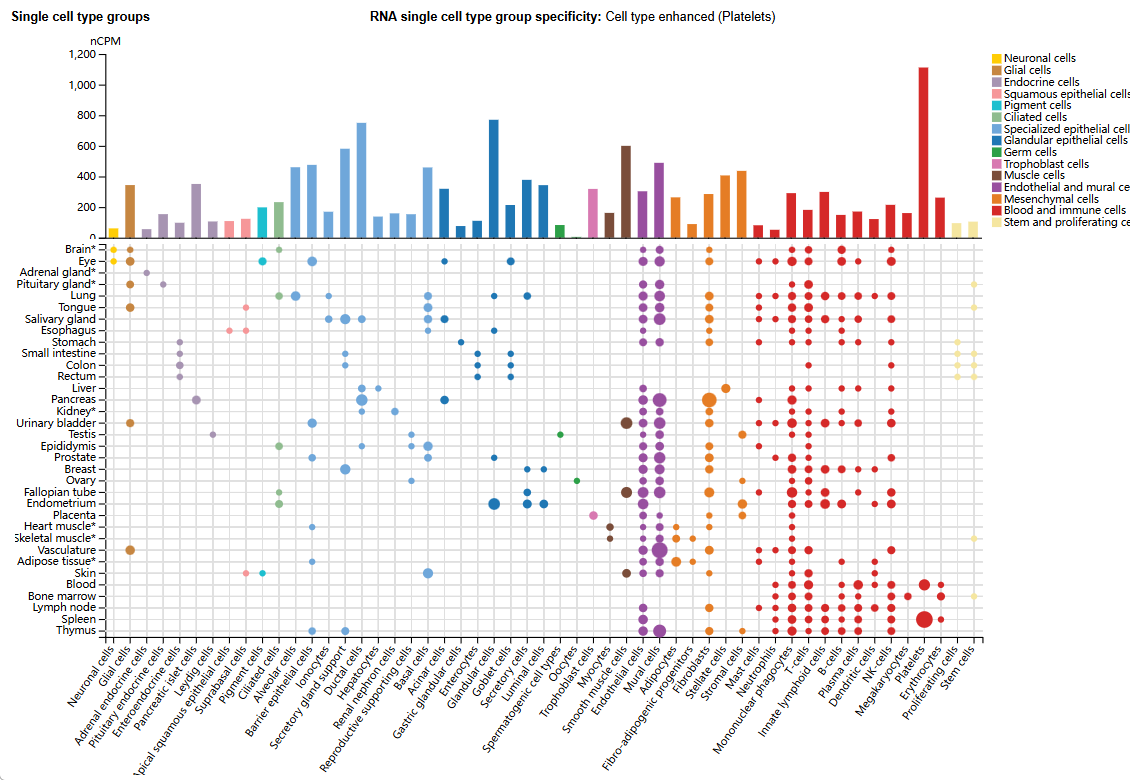
(Data source: uniprot)
ITGA5B1 Structure
The integrin α5 gene (ITGA5) subunit is located on chromosome 12q11. This subunit possesses specific domains, including an extracellular leg domain and a β-propeller domain. These domains are crucial for the function of integrin α5β1, playing a key role, particularly in cell adhesion and signal transduction. The α5 subunit recognizes the arginine-glycine-aspartic acid (RGD) sequence in fibronectin (FN) and fibrinogen. The RGD sequence is a common cell adhesion signal in matrix metalloproteinase (ECM) proteins, especially in ECM proteins such as fibronectin (FN). The binding site for integrin α5β1 to recognize the RGD sequence is formed at the interface between the α5 and β1 subunits, rather than on either subunit alone. This structural arrangement allows α5β1 to specifically recognize and bind to ECM proteins containing the RGD. The extracellular region of the integrin β1 gene (ITGB1) contains a plexin-semaphorin-integrin (PSI) domain, a heterodimer domain, a βI domain containing a metal-dependent adhesion site (MIDAS), and four epidermal growth factor (EGF)-like domains. This gene is located on chromosome 10p11.2. The interaction between integrin α5β1 and its extracellular ligands depends on the MIDAS structure and divalent cations, with calcium ions (Ca²⁺) being a crucial cation for integrin α5β1 ligand binding.
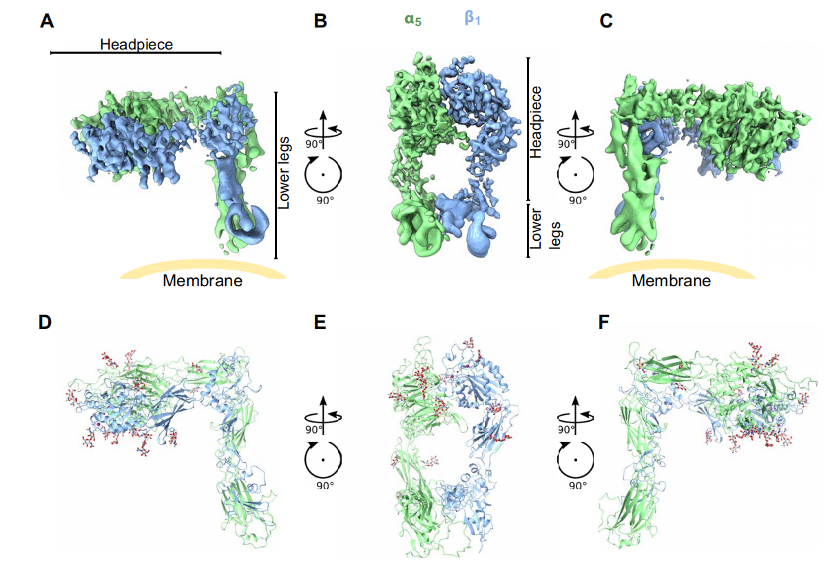
(Data source: Schumacher S, et al. Sci Adv. 2021)
ITGA5B1 signal transduction and modulation functions
ITGA5B1 possesses the typical bidirectional signaling function of integrins. In the "out-to-in" signaling pathway, integrin α5β1 binds to ligands in the extracellular matrix (ECM) and activates downstream signaling pathways, such as MAPK and PI3K, through molecules like FAK and Src. This regulates cell survival, proliferation, adhesion, and migration, as well as the assembly of the ECM. In the "in-to-out" signaling mechanism, activation of integrin α5β1 promotes the regulation of interactions between the cell and the ECM. Intracellular calcium levels, PKC, TGF-β signaling, and key proteins such as Talin and Kindlin bind to the β1 tail of integrin, activating conformational changes in integrin α5β1.
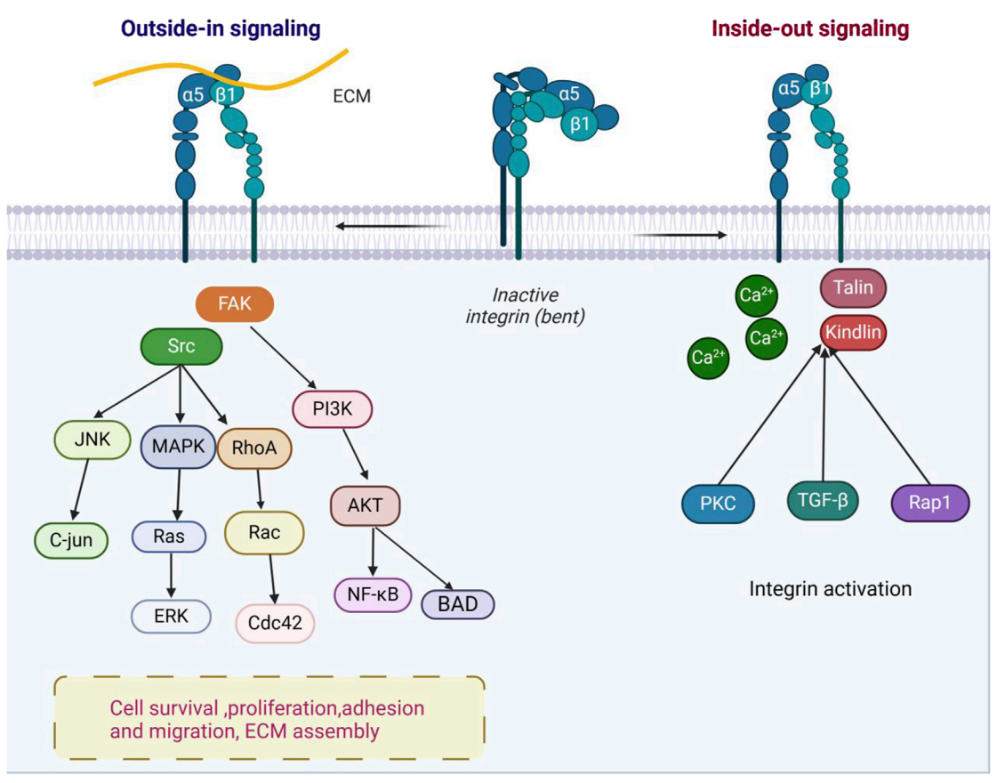
(Data source: Li X, et al. Front Cell Dev Biol. 2025)
Osteogenesis is regulated by integrin α5β1. This process begins with the differentiation of mesenchymal stromal cells (MSCs) into pre-osteoblasts, which subsequently mature into fully functional osteoblasts. During this process, α5β1 is crucial for promoting osteoblast adhesion, lineage specialization, and responses to the external matrix. Mature osteoblasts secrete bone matrix proteins to drive new bone formation and undergo programmed apoptosis after completing their synthetic function. α5β1 binds to extramammary ligands containing RGD domains, promoting osteoblast adhesion and migration. Furthermore, osteoogenesis is also regulated by fibroblast growth factor, bone morphogenetic proteins, hormones, extramammary matrix components, and mechanotransmission; these factors collectively act on the regulation of α5β1, thereby finely modulating bone formation.
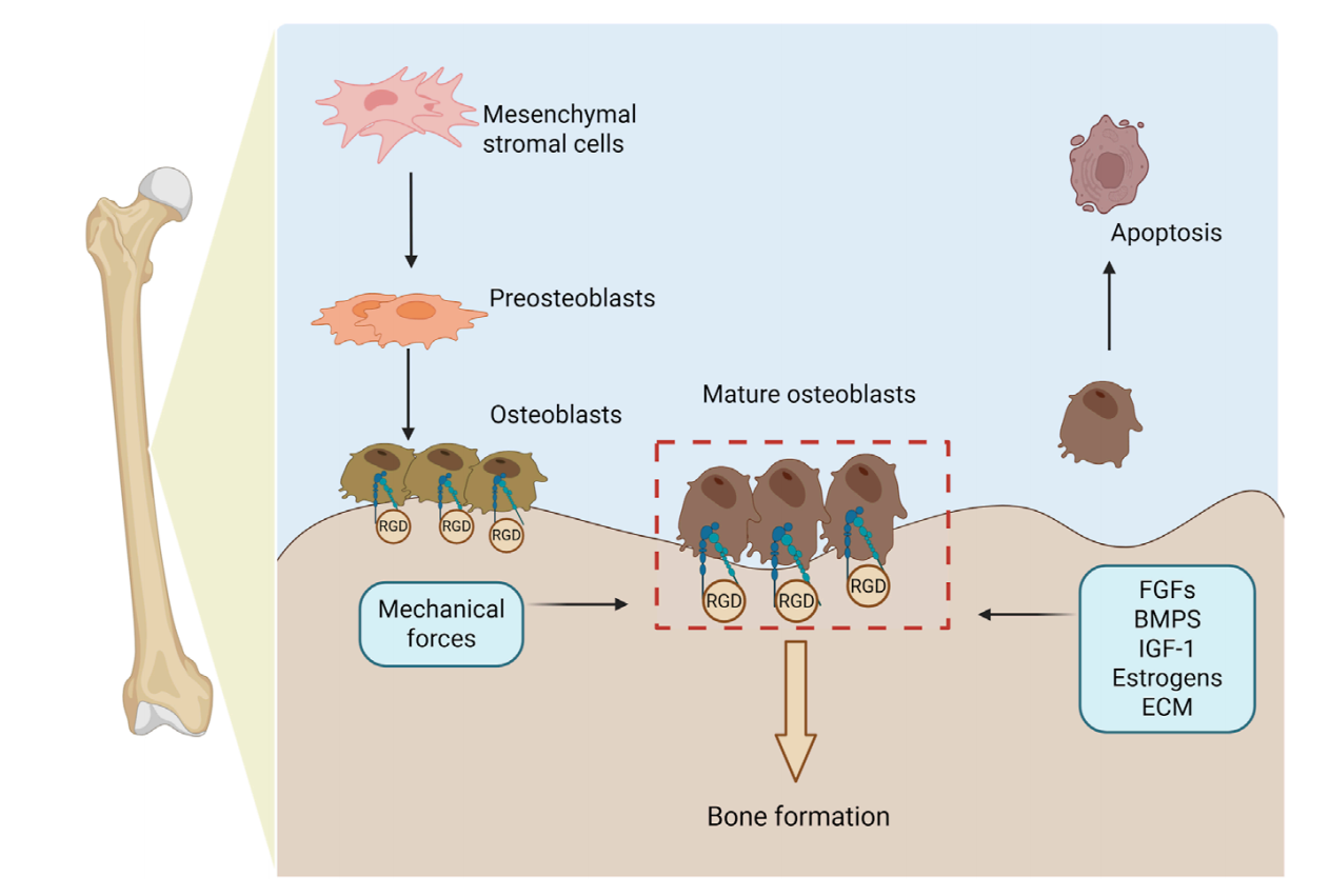
(Data source: Li X, et al. Front Cell Dev Biol. 2025)
The role of ITGA5B1 in disease
In osteoarthritis, damage to the cartilage matrix can lead to the production of inflammatory cytokines, matrix metalloproteinases, and fibronectin fragments, which degrade collagen. Fibronectin fragments bind to α5β1 and TLR, activating the NF-κB and MAPK signaling pathways, further releasing inflammatory mediators and MMPs, resulting in widespread matrix destruction. Simultaneously, the binding of α5β1 to immune cells can induce the expression of IL-1β, TNF-α, and MMPs, exacerbating synovial inflammation and cartilage degeneration. This α5β1-driven signaling pathway also plays a role in bone metastasis, accelerating tumor-induced bone destruction.
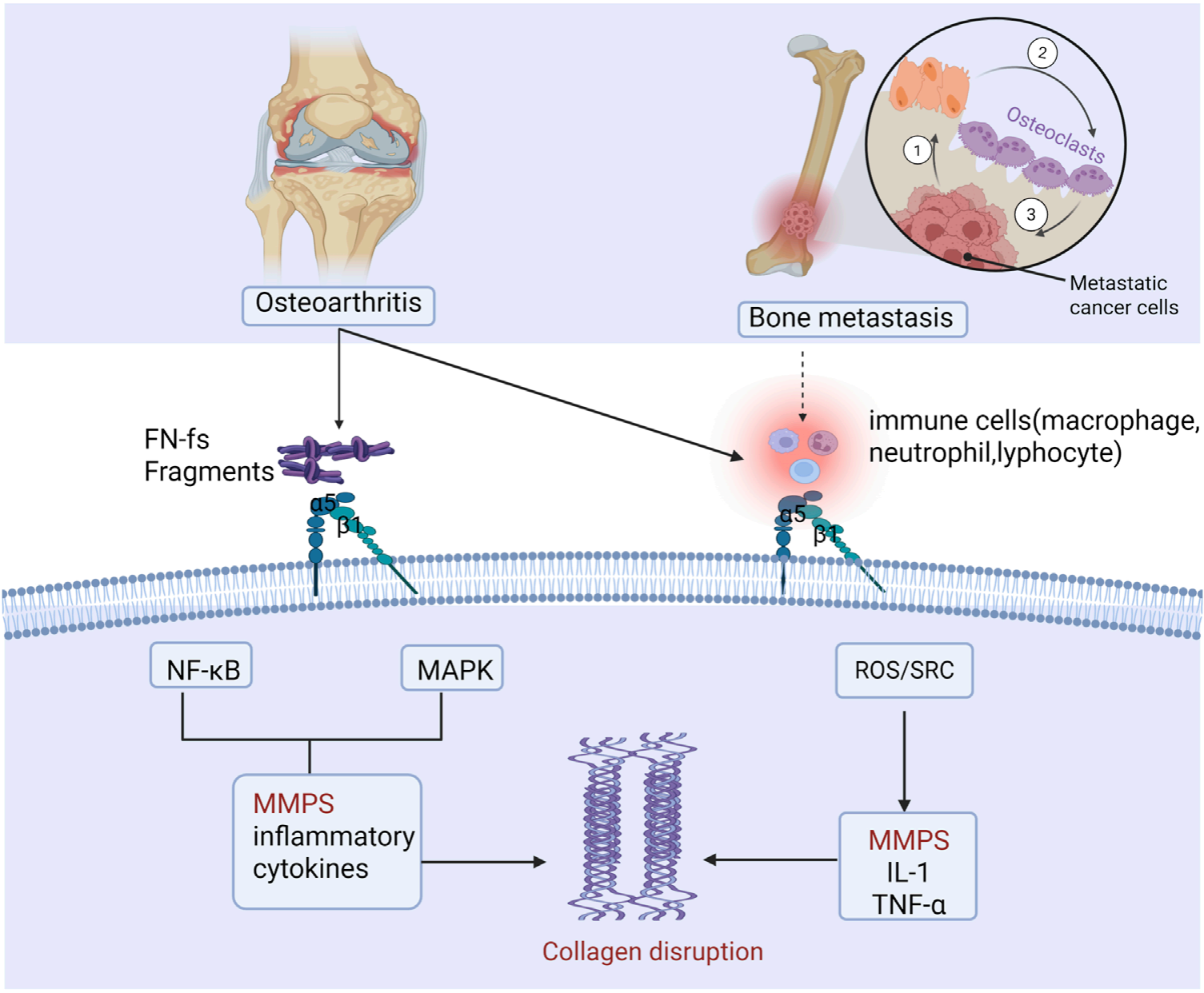
(Data source: Li X, et al. Front Cell Dev Biol. 2025)
Integrin α5β1 plays different functions in different cellular compartments of the PDAC tumor microenvironment. In tumor cells, α5β1 promotes fibronectin (FN)-dependent adhesion, focal adhesion/Src signaling, proliferation, invasion, and treatment resistance. In pancreatic stellate cells (PSCs)/cancer-associated fibroblasts (CAFs), α5β1 signaling drives fibroblast activation, extracellular matrix remodeling, fibrosis, and tumor-matrix interactions, promoting tumor cell invasion, hypoxia, and treatment resistance. In endothelial cells, FN-α5β1 signaling regulates endothelial cell migration, angiogenesis, and vascular remodeling.
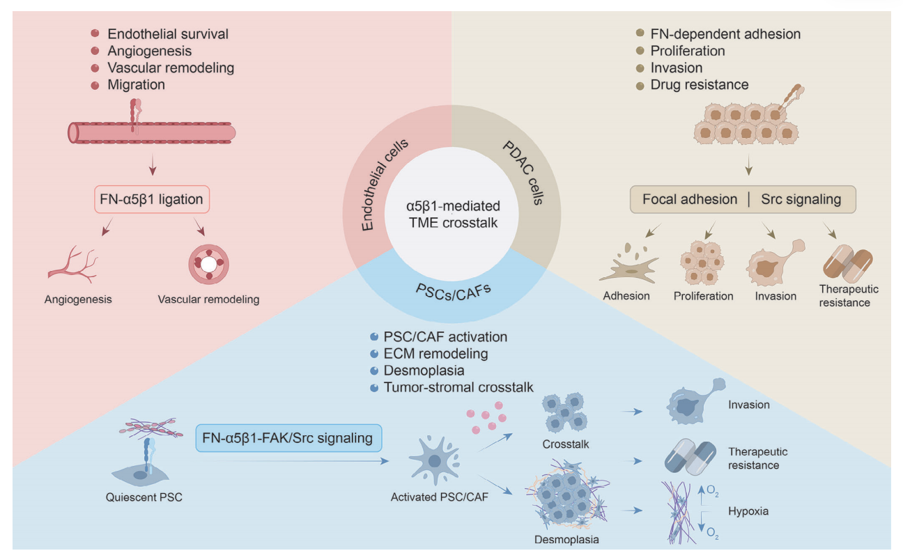
(Data source: Ma C, et al. Clin Transl Med. 2026)
ITGA5B1 targeted therapy
Integrin α5β1 plays a crucial role in mediating tumor cell adhesion, migration, survival, and tumor-matrix interactions, making its targeting a promising strategy in cancer therapy. Integrin α5β1 primarily serves as the main receptor for fibrinogen (FN) in the extracellular matrix. Its activation triggers downstream signaling pathways such as FAK, PI3K/AKT, and MAPK/ERK, which collectively promote tumor progression, invasion, and treatment resistance. In pancreatic ductal adenocarcinoma, the FN-α5β1 axis is closely associated with the fibrotic tumor microenvironment, where mesenchymal stem cells produce abundant extracellular matrix components, thereby enhancing integrin-mediated signal transduction. Therefore, targeting integrin α5β1 may simultaneously inhibit intrinsic tumor cell signaling and disrupt tumor-matrix crosstalk, offering a dual therapeutic advantage.
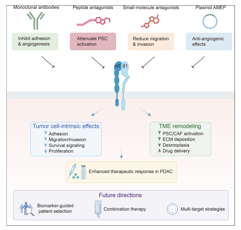
Volociximab is a chimeric monoclonal antibody that targets α5β1, blocking the interaction between α5β1 and fibronectin (FN), thereby inhibiting integrin-mediated cell adhesion and angiogenesis. In a multicenter, open-label phase II trial, Volociximab, in combination with gemcitabine, achieved partial remission in 1 of the 20 reported patients, with 10 having stable disease and a median overall survival of 5.4 months. Although this combination regimen was considered feasible, the low objective response rate and single-arm design limited definitive assessment of the additional clinical benefit of Volociximab. In platinum-resistant epithelial ovarian cancer, Volociximab monotherapy was generally well-tolerated, but its antitumor efficacy was limited. In advanced non-small cell lung cancer (NSCLC), its combination therapy with cisplatin and paclitaxel showed manageable toxicity and preliminary clinical activity. Early clinical trials of Volociximab showed feasibility, but its antitumor activity was only mild and dependent.
PF-04605412 is a next-generation therapeutic antibody with dual functionalities. This fully human IgG1 monoclonal antibody is designed not only to block the interaction between integrin α5β1 and FN, but also to induce antibody-dependent cytotoxicity (ADCC). Preclinical studies have shown that PF-04605412 effectively binds to integrin α5β1 on tumor cells and triggers immune-mediated cytotoxicity, thereby enhancing antitumor activity beyond simple integrin blockade. In a first-in-human Phase I clinical trial, PF-04605412 showed evidence of target binding and acceptable pharmacokinetic properties. However, infusion-related reactions were observed, and overall antitumor activity was limited, ultimately restricting further clinical development of this antibody.
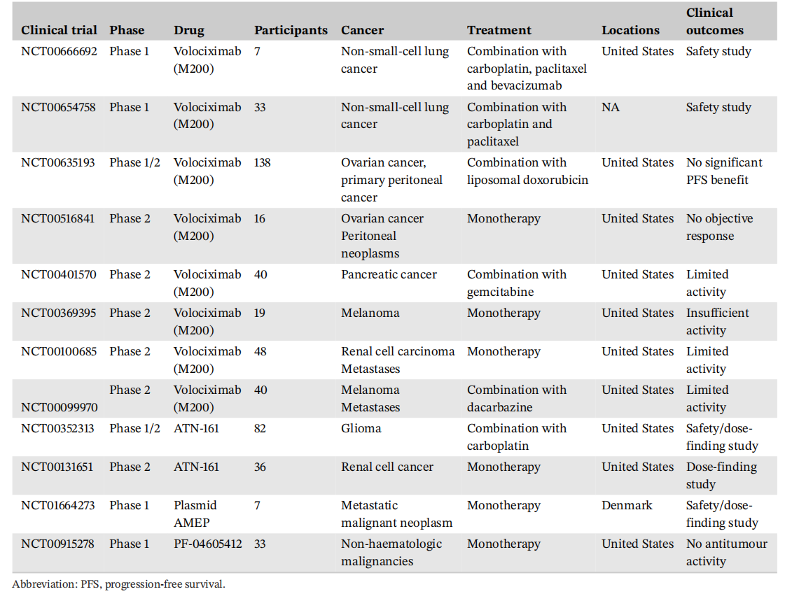
(Data source: Ma C, et al. Clin Transl Med. 2026)
