Mannoceramide GM3 is a fundamental glycolipid on the surface of mammalian cells and a key regulator of transmembrane signaling and cell recognition. In oncology, GM3 exerts its tumor-suppressive effect by regulating the activity of various receptor tyrosine kinases (RTKs) and their downstream pathways. N-hydroxyacetylneuraminic acid GM3 ganglioside (NeuGcGM3), also known as NGcGM3, is a monosialotetrahexosylganglioside belonging to the cell membrane surface glycosphingolipids. It is almost not expressed in normal human tissues but functions as a highly specific neoantigen in various solid tumors.
Structure of GM3 and NeuGcGM3
Ceramide glycolipids (GM3) are sialic acid-containing glycolipids distributed in the plasma membranes of vertebrates, mediating cell signaling and membrane organization. Structurally, GM3 consists of a hydrophobic ceramide moiety anchored in the outer lamina of the lipid bilayer, and a hydrophilic oligosaccharide head extending into the extracellular space. The biological function of GM3 is influenced by its structural heterogeneity, which stems from variations in the sphingosine moiety and sialic acid residues . There are two variants of GM3, both possessing a conserved ceramide and lactose core. They branch at the terminal sialic acid level and are defined as N- acetylneuraminic acid (Neu5Ac) or N- hydroxyacetylneuraminic acid (Neu5Gc).
NeuGcGM3 belongs to the monosialotetarian ganglioside family and is one of the simplest gangliosides in terms of structure. Its molecule consists of three parts.
Hydrophobic ceramide anchoring region: embedded in the cell membrane lipid bilayer, fixing molecules to the outer leaflet of the plasma membrane.
Hydrophilic oligosaccharide head group: composed of glucose (Glc), galactose (Gal) and sialic acid, extending into the extracellular space.
Key structural difference: Unlike NeuAcGM3 (containing N-acetylneuraminic acid) expressed by normal human cells, NeuGcGM3 has N-hydroxyacetylneuraminic acid residues (NeuGc/Neu5Gc), which means that there is an extra hydroxyl group (-OH) on the N-acetyl group.
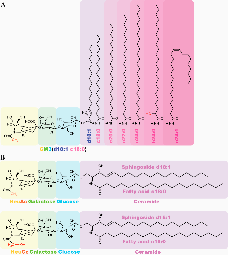
(Data source: Zeng M, et al. Biomolecules. 2026)
The role of NeuGcGM3 in tumor progression and the immune system
NeuGcGM3 glycolipids are expressed on the surface of tumor cells, typically located within lipid rafts. Interactions between this glycolipid and other lipid raft components, such as EGFR, may enhance signal transduction via this receptor. Pathological accumulation of Neu5GcGM3 on the cell surface is driven by the metabolic incorporation of exogenous n-hydroxyacetylneuraminic acid and a deficiency of the enzyme responsible for Neu5AcGM3 synthesis. This altered sialylation promotes hyperphosphorylation of rtk (including EGFR, PDGFR, and FGFR), ultimately accelerating malignant transformation and tumor development.
This glycolipid, released into the tumor microenvironment, has an immunosuppressive effect. Its interaction with CD4+ T lymphocytes prompts insertion of the glycolipid into the cell membranes, leading to downregulation of CD4 molecules. The shedding of NeuGcGM3 glycolipid also interferes with dendritic cell differentiation and maturation, resulting in a reduction in the number of these cells.
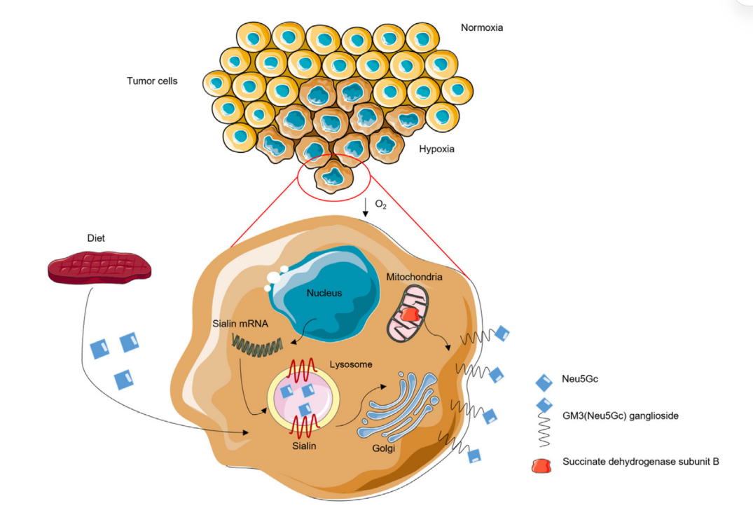
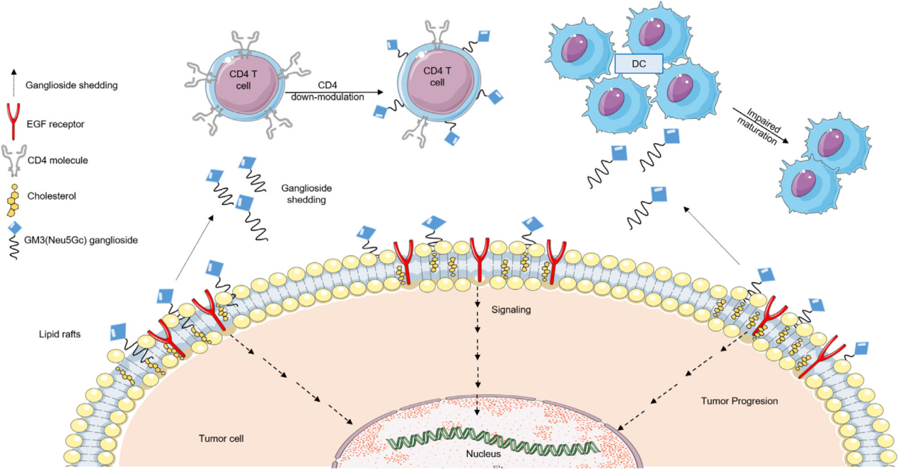
(Data source: Labrada M, et al. Semin Oncol. 2018)
NeuGcGM3 targeted therapy
Racotumomab is an anti-heterogeneous monoclonal antibody vaccine targeting NeuGcGM3 ganglioside, a tumor-associated antigen expressed on the surface of various cancer cells, including non-small cell lung cancer. By mimicking this antigen, Racotumomab prompts the immune system to recognize and attack cells carrying NeuGcGM3, thereby inhibiting tumor growth.
Chimeric antigen receptor (CAR) T cells engineered to express single-chain variable fragments targeting NeuGcGM3 (e.g., fragments derived from 14F7 antibodies) have successfully controlled NeuGcGM3-positive ovarian xenografts in vivo without causing off-target toxicity. This NeuGcGM3-targeting strategy has also been integrated into innovative delivery platforms, such as artificial cell-derived vesicles (ACDVs) derived from cytotoxic T cells and NK92 cells. Conjugating these vesicles to anti-NeuGcGM3 antibodies (such as 14F7hT) significantly enhanced tumor cell interactions, endocytosis, and targeted cytotoxicity.
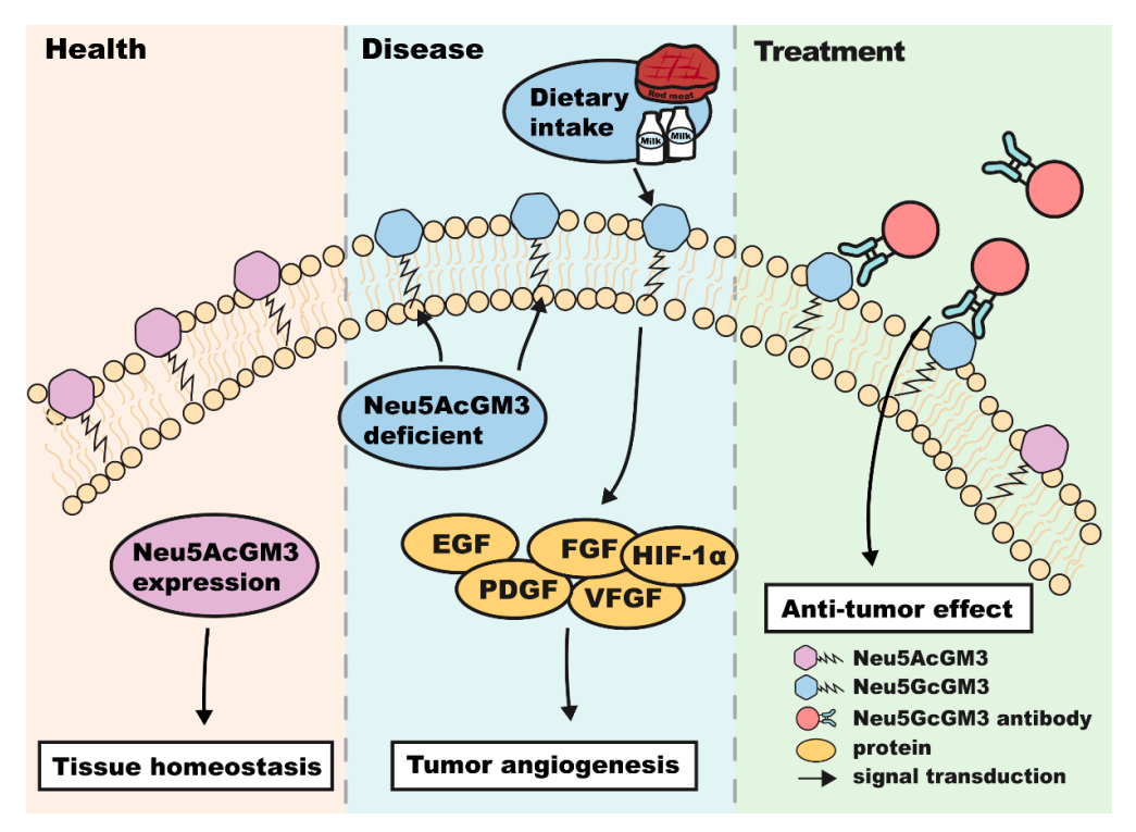
(Data source: Zeng M, et al. Biomolecules. 2026)
