Service Support
Mabnus Biotech provided customized ATG9A phosphorylated antibody services and offered Flag antibody affinity gel products.
Background
Chemotherapy, used alone or in combination with immunotherapy, is a routine approach in cancer treatment. Autophagy enables cancer cells to tolerate metabolic and therapeutic stress, leading to resistance to chemotherapy, targeted therapy, and immunotherapy. Autophagy is a key factor determining the effectiveness of chemotherapy.
A key factor and a potential therapeutic target for overcoming drug resistance, the cyclic GMP-AMP synthase (cGAS)-interferon gene stimulating factor (STING) pathway, initially identified as a cytoplasmic DNA sensing system that initiates innate immune responses, has recently become a key regulator in cancer biology. However, whether the cGAS - STING pathway crosstalks with autophagy, and how this crosstalk affects chemotherapy resistance, remains poorly understood.
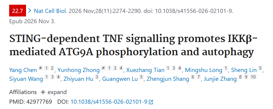
In 2026, Zhang Junjie's team at the Institute of Medical Sciences, Wuhan University, published a study titled "STING-dependent TNF signaling promotes IKKβ-mediated ATG9A phosphorylation and autophagy." Using head and neck squamous cell carcinoma as a model, this study found that chemotherapy can activate the cGAS-STING pathway, thereby inducing TNF expression. TNF mediates paracrine signaling in receptor tumor cells, leading to IKKβ activation and ATG9A phosphorylation at serine 654, thus promoting autophagy. Furthermore, TNF, nutrient deprivation, and mTOR inhibition can all induce IKKβ activation and ATG9A phosphorylation at S654, suggesting that the IKKβ-ATG9A axis may be a common regulatory mechanism for autophagy. Analysis of patient samples further showed that chemotherapy can induce autophagy, and increased ATG9A phosphorylation at S654 is associated with poor chemotherapy response. Blocking TNF can enhance the efficacy of chemotherapy alone and chemotherapy combined with immune checkpoint inhibitory therapy. The findings collectively reveal a previously unknown TNF-IKKβ-ATG9A signaling axis that promotes autophagy and highlight that blocking TNF is a promising strategy for overcoming chemotherapy resistance.
Chemotherapy can induce autophagy in HNSCC
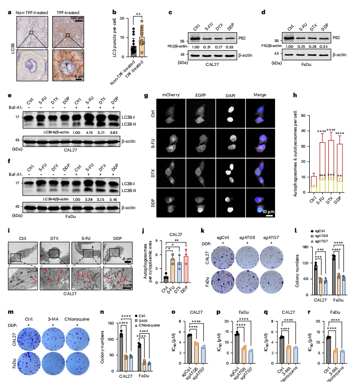
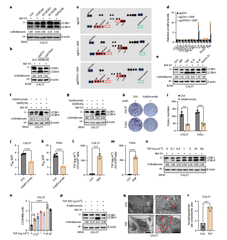
TPF regimens (DTX, DDP, and 5-FU) are first-line treatments for HNSCC and have been shown to reduce metastasis and improve patient survival. Analysis of tissue samples from HNSCC patients revealed that cancer cells in patients receiving chemotherapy exhibited significantly higher levels of autophagy compared to untreated patients, as evidenced by increased LC3 dot staining. Treatment of two HNSCC cell lines, CAL27 and FaDu, with TPF drugs alone reduced p62 levels. All three chemotherapeutic agents significantly enhanced LC3B-II accumulation after Baf-A1 treatment . Dual-fluorescence LC3 reporter system analysis showed an increase in both autophagosomes and autolysosomes, and transmission electron microscopy revealed increased autophagosomes, confirming enhanced autophagic flux. These data suggest that chemotherapy activates autophagy in HNSCC, thereby promoting chemotherapeutic resistance.
STING–NF-κB axis is essential for chemotherapy-induced autophagy
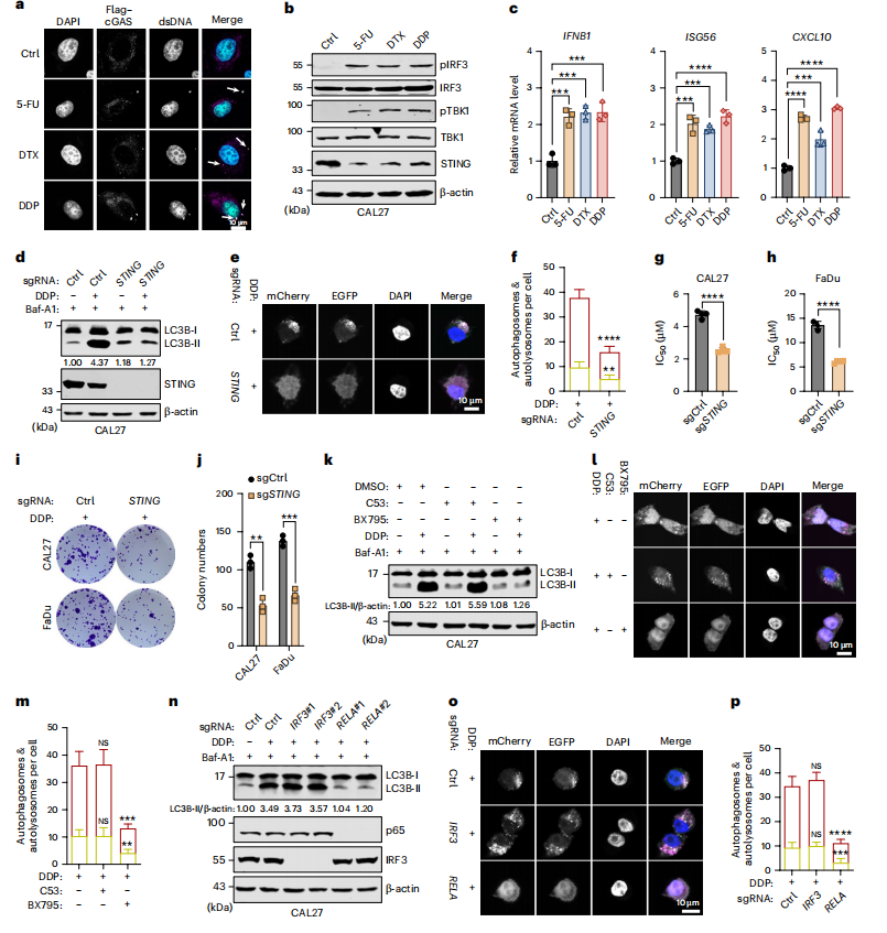
Chemotherapy-induced DNA damage is a major driver of cytotoxicity, leading to the accumulation of double-stranded DNA (dsDNA) in the cytoplasm, which co-localizes with cGAS. This genomic toxicity is accompanied by strong activation of the cGAS-STING pathway, manifested as STING degradation, phosphorylation of TBK1 and IRF3, and upregulation of downstream genes such as IFNB1, ISG56, and CXCL10. STING gene knockout eliminated DDP-induced LC3B-II accumulation and autophagosome formation , indicating that chemotherapy-induced autophagy depends on the intact cGAS-STING pathway. STING deficiency significantly enhanced the sensitivity of HNSCC cells to chemotherapy. Chemotherapy-induced DNA damage activates the cGAS-STING-TBK-NF-κB axis, thereby promoting autophagy and enhancing chemotherapy resistance.
TNF -mediated paracrine induction of autophagy promotes chemotherapy resistance
Activation of NF-κB promotes the secretion of inflammatory cytokines such as TNF, IL-1β, and IL-6. Conditioned culture medium (CM) transfer experiments showed that CM collected from chemotherapeutic donor cells significantly induced LC3B-II accumulation and increased the formation of autophagosomes and autolysosomes in recipient cells. This indicates that chemotherapeutic cells release soluble factors that promote autophagy in recipient cells. DDP treatment significantly increased the secretion of MIF, TNF, and IL-6, while IL-8 and Serpin E1 were decreased after STING knockout. TNF is the main mediator of paracrine induction of autophagy after chemotherapy. Functionally, adalimumab blocking of TNF significantly improved the sensitivity of HNSCC cells to chemotherapy, as evidenced by impaired colony formation survival and a decreased IC50 value. TEM further confirmed that TNF treatment significantly increased the number of autophagic vesicles. These data suggest that chemotherapy-induced TNF promotes autophagy in untreated recipient cells through a paracrine mechanism, leading to chemotherapy resistance; while TNF blockade can effectively enhance chemosensitivity.
TNF blockade can enhance the therapeutic effects of chemotherapy alone and chemotherapy combined with PD-1 inhibition
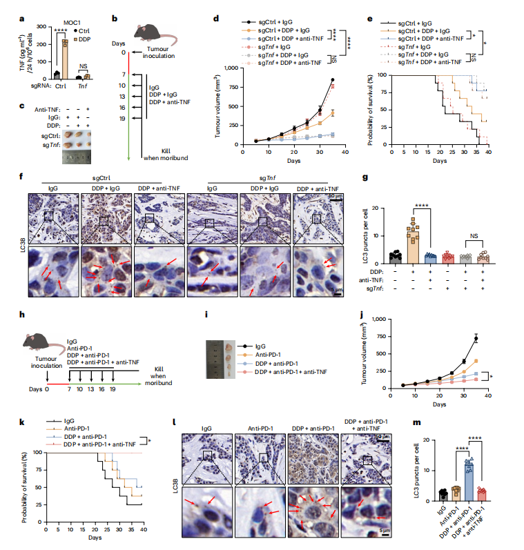
DDP treatment significantly increased TNF secretion in control MOC1 cells, while this response was almost completely eliminated in TNF- knockout MOC1 cells. In the absence of chemotherapy, TNF deficiency had little effect on tumor growth. However, after DDP treatment, TNF- knockout tumors showed significantly slower growth and improved survival compared to control tumors. Similarly, DDP significantly increased LC3 spot formation in control tumors, an effect almost entirely absent in TNF- knockout tumors. Furthermore, in mice carrying TNF- knockout tumors, TNF neutralization did not further inhibit tumor growth, improve survival, or reduce LC3 spots. These results suggest that tumor cell-derived TNF plays a crucial role in this model, promoting chemotherapy-induced protective autophagy and chemotherapeutic resistance.
When used in combination with DDP and anti-PD-1 therapy, TNF neutralization further inhibited tumor growth and improved survival in mice. Immunohistochemical (IHC) analysis further showed that TNF neutralization significantly reduced the formation of LC3 spots in tumors treated with DDP and anti-PD-1. These data collectively suggest that TNF produced by tumor cells promotes protective autophagy and enhances drug resistance, while blocking TNF can improve the efficacy of chemotherapy alone and in combination with PD-1 inhibitors.
TNF-induced autophagy requires IKKβ
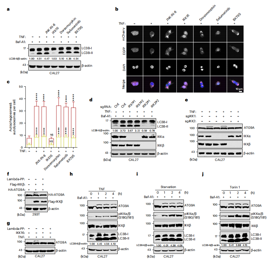
Only the IKK16 inhibitor significantly suppressed TNF-induced LC3B-II accumulation and autophagosome formation. Under TNF treatment, the absence of IKKβ but the lack of IKKα completely inhibited autophagy induction, indicating that IKKβ is a key downstream kinase. ATG9A is indispensable for TNF-induced autophagy, as TNF failed to promote LC3B-II accumulation in ATG9A knockout cells. Furthermore, ATG9A exhibited a significant migration shift after TNF treatment, consistent with its underlying phosphorylation. This band shift was eliminated in IKKβ-deficient cells but remained intact in IKKα-reduced cells. These data suggest that TNF-stimulated IKKβ activation is crucial for autophagy and indicate that IKKβ-mediated ATG9A phosphorylation is a co-regulatory event in the multi-stimulatory-induced autophagy process.
IKKβ phosphorylates ATG9A at position S654 to promote autophagy
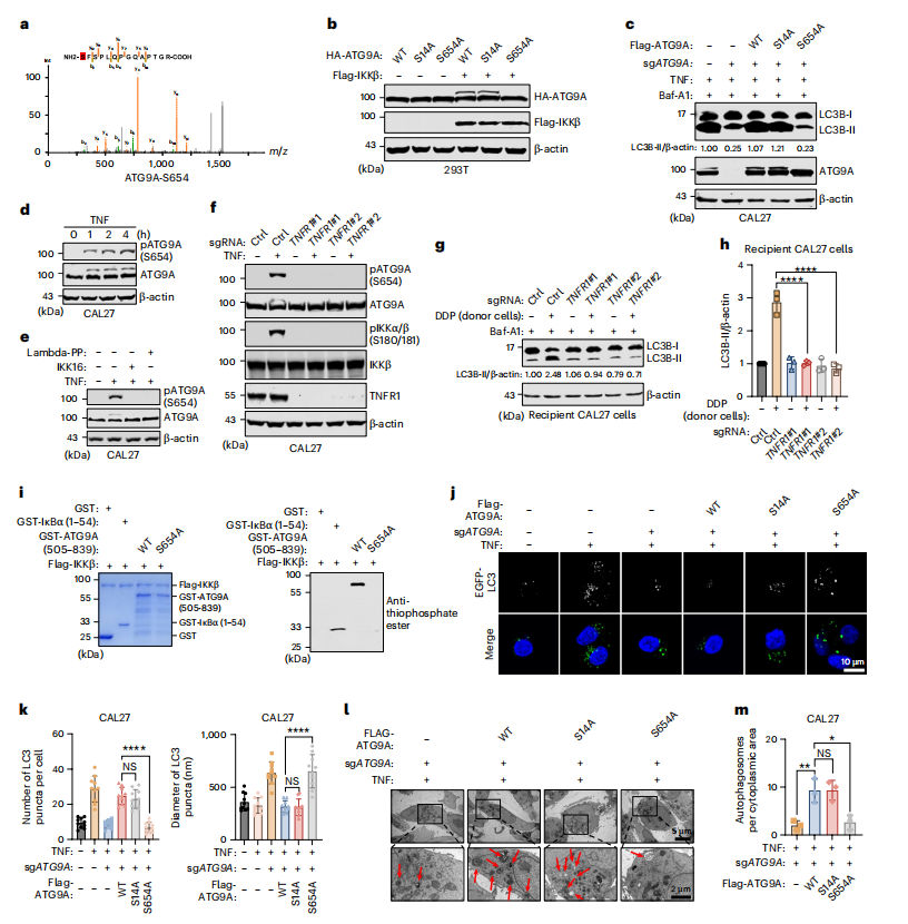
Immunoprecipitation of endogenous ATG9A, followed by phosphoproteomic analysis, identified serine 14 (S14) and serine 654 (S654) as candidate phosphorylation sites. These sites were strongly induced by TNF but showed a marked reduction in the absence of IKKβ. The S654A mutation completely abolished TNF-induced ATG9A translocation, whereas the S14A mutation had no such effect, establishing S654 as the specific site phosphorylated by IKKβ. Reconstitution of ATG9A-knockout cells with the S654A mutant—but not the S14A mutant—failed to restore TNF-induced LC3B-II accumulation. A phospho-specific antibody targeting p-ATG9A (S654) was generated, and its specificity was validated through three independent experimental approaches. Using this antibody, it was observed that TNF treatment rapidly induced phosphorylation of ATG9A at S654, a process that could be abolished by treatment with λ-phosphatase or the IKK inhibitor IKK16.
In vitro kinase assays demonstrated that purified IKKβ can directly phosphorylate the cytoplasmic C-terminal domain (amino acids 505–839) of recombinant ATG9A; this effect was absent in the S654A mutant, confirming that IKKβ directly phosphorylates ATG9A at the S654 site. Transmission electron microscopy (TEM) analysis confirmed that the S654A mutant fails to support autophagosome formation upon TNF stimulation, while a phosphomimetic S654D mutation restores autophagy and bypasses the requirement for IKKβ. Furthermore, the S654A mutation sensitizes cells to chemotherapy. These findings indicate that IKKβ promotes autophagy by phosphorylating ATG9A at S654, thereby contributing to chemotherapy resistance.
IKKβ-mediated phosphorylation of ATG9A at position S654 enhances lipid dephospholipase activity and autophagy fuction
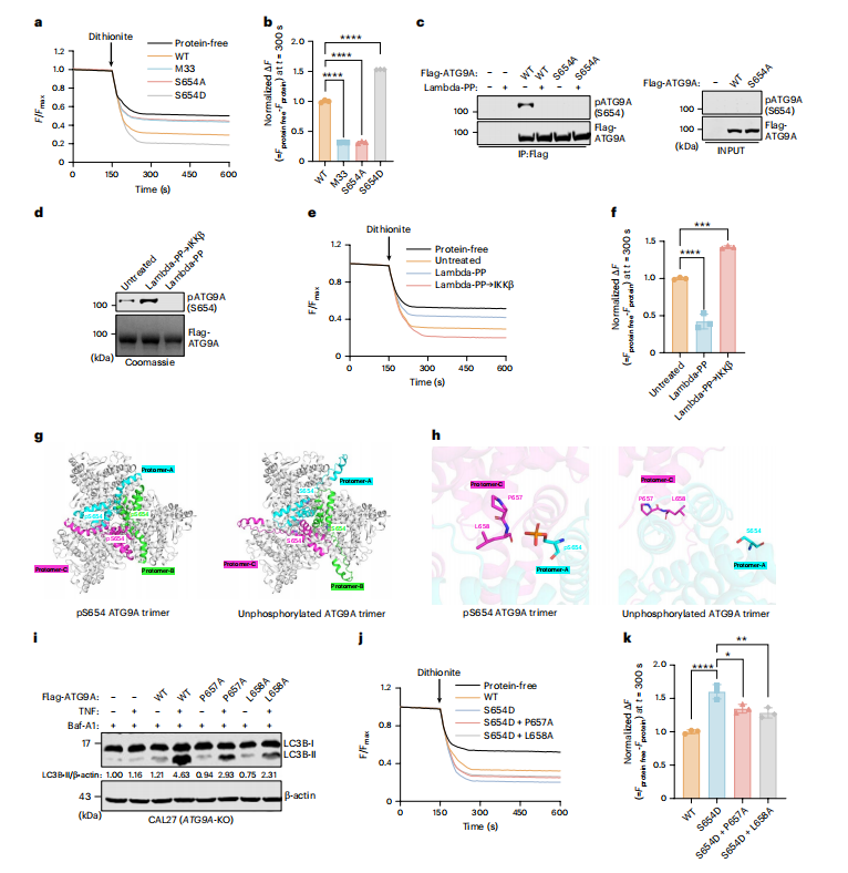
In in vitro lipid scrambling experiments, the S654A mutation significantly reduced the lipid scrambling enzyme activity, while the S654D mutation significantly enhanced it. λ phosphatase treatment significantly reduced the lipid scrambling enzyme activity of ATG9A, while subsequent in vitro phosphorylation via IKKβ significantly enhanced this activity. This indicates that IKKβ-mediated phosphorylation of ATG9A at position S654 can enhance its lipid scrambling enzyme activity.
AlphaFold 3 predicts that phosphorylation of S654 may promote a unique interprotein body arrangement in the C-terminal region, thereby facilitating the formation of a flipper-like conformation. The P657-L658 region was found to be functionally essential in the S654 phosphorylation-mediated enhancement of ATG9A lipid flipper activity.
These data suggest that IKKβ-mediated phosphorylation of ATG9A at position S654 promotes its unique interprotein arrangement within the flexible C-terminal region, thereby enhancing lipid deordering enzyme activity and autophagy, which in turn contributes to the development of chemical resistance.
TNF blockade overcomes chemotherapy resistance by inhibiting autophagy flux driven by ATG9A S654 phosphorylation
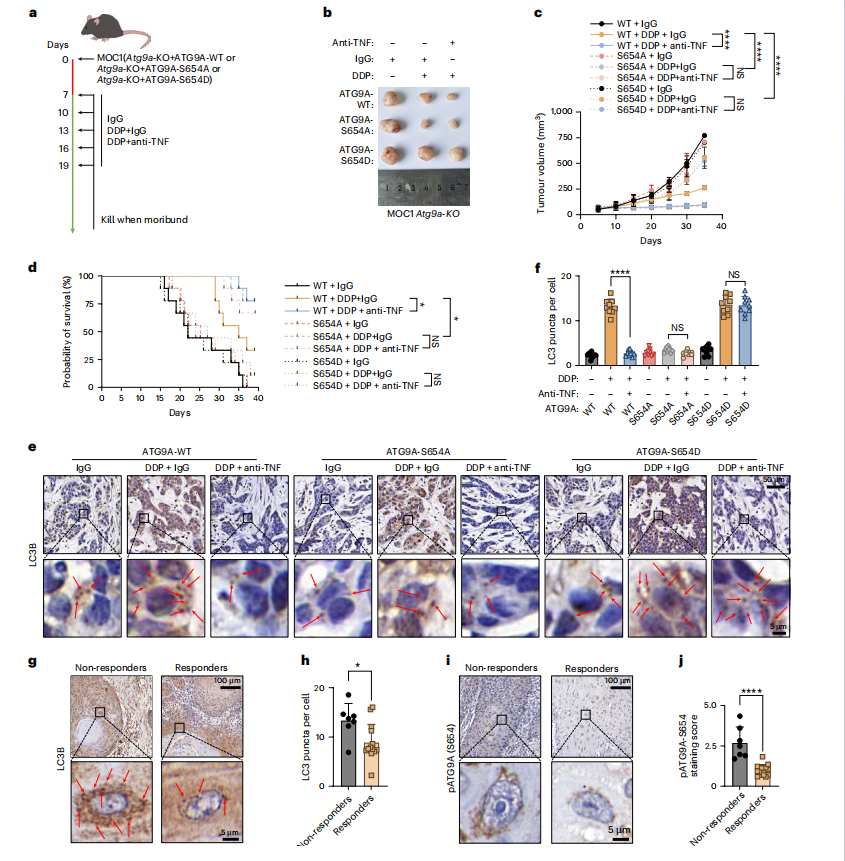
MOC1 Atg9a knockout cells were reintroduced into homologous mice carrying wild-type ATG9A, phosphate-deficient S654A mutants, or phosphate-mimicking S654D mutants. In mice carrying wild-type ATG9A tumors, TNF neutralization significantly enhanced the antitumor effect of DDP and prolonged survival. In contrast, S654A tumors were already more sensitive to DDP, and anti-TNF treatment did not provide additional therapeutic benefit. Conversely, S654D tumors remained relatively resistant to DDP, and TNF neutralization did not further enhance the sensitivity of these tumors to DDP. S654A tumors showed lower LC3 spot formation after DDP treatment, regardless of TNF blockade; while S654D tumors maintained higher LC3 spot levels despite anti- TNF treatment. These findings suggest that TNF blockade enhances the efficacy of chemotherapy by preventing ATG9AS654 phosphorylation and the resulting protective autophagy. TNF blockade overcomes chemotherapy resistance by inhibiting autophagic flux driven by ATG9A S654 phosphorylation, and p-ATG9A (S654) levels are positively correlated with chemotherapy resistance.
Summary
This study demonstrates that chemotherapy activates the cGAS-STING signaling pathway, thereby inducing TNF secretion, which in turn triggers IKKβ-mediated phosphorylation of ATG9A at serine 654, enhancing its lipid scrambling activity, triggering protective autophagy, and promoting chemoresistance. These findings establish inflammatory paracrine signaling as an important mechanism for tumor adaptation to chemotherapy and indicate that TNF blockade is a promising strategy for overcoming chemoresistance.
